

 微信在线咨询


| 产品编号 | 产品名称 | 产品包装 | 产品价格 |
| P2206S | HA标签蛋白免疫沉淀试剂盒(琼脂糖凝胶法) | 20次 | 817.00元 |
| P2206M | HA标签蛋白免疫沉淀试剂盒(琼脂糖凝胶法) | 100次 | 3106.00元 |
碧云天生产的HA标签蛋白免疫沉淀试剂盒(琼脂糖凝胶法) (HA-tag Protein IP Assay Kit with Agarose Gel)是一种通过高特异性的Anti-HA琼脂糖凝胶进行HA标签融合蛋白免疫沉淀或免疫共沉淀的试剂盒。本产品的免疫沉淀产物,可以用于HA标签融合蛋白或其蛋白复合物组分的检测。
本试剂盒包含高质量的Anti-HA琼脂糖凝胶及经过优化验证的免疫沉淀必要试剂,使免疫沉淀(Immunoprecipitation, IP,也称Pull-down)或免疫共沉淀(Co-IP)实验更加简单、便捷、高效,广泛用于带有HA标签的融合蛋白或其蛋白复合物的免疫沉淀、免疫共沉淀或纯化等实验。
免疫沉淀或免疫共沉淀是研究蛋白或蛋白与蛋白相互作用(Protein-Protein Interactions, PPIs)的常用实验技术,通过使用特异性抗体和可结合抗体的介质(如Protein A/G Agarose或Protein A/G磁珠),或直接使用偶联特异性抗体的介质(如琼脂糖凝胶或磁珠),然后通过离心或磁力从溶液中分离出抗原和抗体复合物,从而将目标蛋白质从复杂样品中分离出来,随后可以用于Western印迹检测或质谱分析等[1-2]。
HA标签(HA-tag)、Myc标签(Myc-tag)、Flag标签(Flag-tag)、V5标签(V5-tag)、His标签(His-tag)和GST标签(GST-tag)等是表达载体上最常见的一些标签,通过与这些标签的融合表达可以非常方便地检测目的蛋白及与目的蛋白相互结合的蛋白,也可以非常方便地用于目的蛋白的纯化。
HA-tag是9个氨基酸残基(YPYDVPDYA)组成的多肽,源于人流行性感冒病毒血凝素(Influenza virus hemagglutinin)的98-106位氨基酸序列,所以称为HA多肽。其常用的形式有HA和3X HA,通过基因重组技术把HA-tag的核酸序列与目的基因的5'端或3'端连接,就可以最终表达形成HA-tag的目的蛋白。HA-tag具有以下优点:HA-tag通常不会与目的蛋白相互作用,并且大多数情况下不会影响目的蛋白的功能;HA-tag作为标签蛋白,后续通过HA抗体(AH158/AF2858)、Anti-HA磁珠(P2121)和Anti-HA凝胶(P2287)即可对目的基因的表达、定位及功能进行检测或对目的蛋白进行纯化、免疫沉淀或免疫共沉淀等。基于以上优点,HA标签已被广泛应用于蛋白表达、纯化、鉴定、相互作用和功能等多方面的研究[3]。
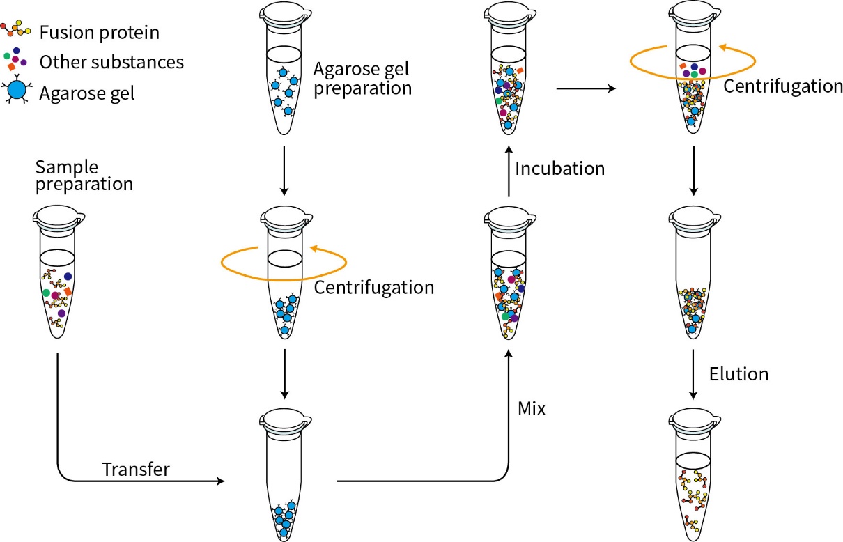
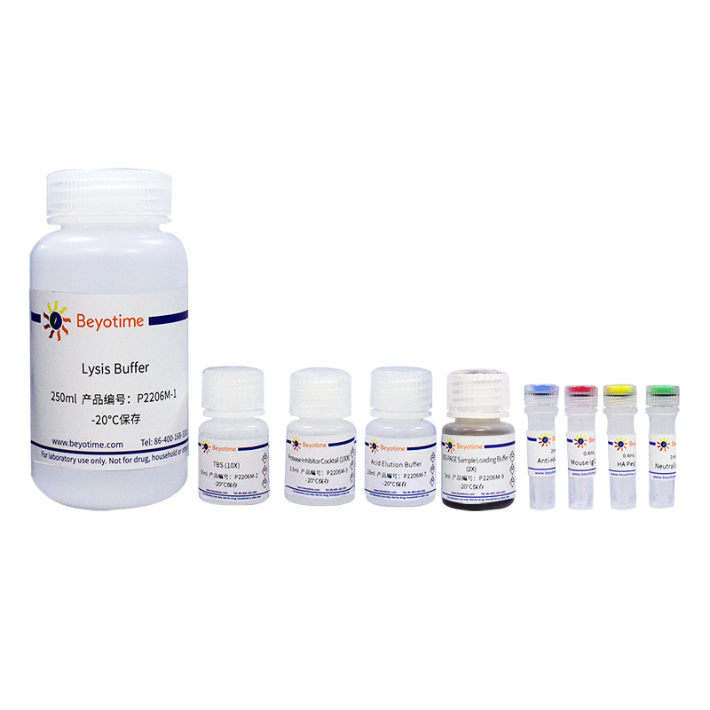
本试剂盒包含高质量的Anti-HA Agarose Gel (Anti-HA琼脂糖凝胶)、Mouse IgG Agarose Gel (小鼠IgG琼脂糖凝胶,作为阴性对照)及优化的各种缓冲液如Lysis Buffer、TBS (10X)、Protease Inhibitor Cocktail (100X)、HA Peptide (25X)、Acid Elution Buffer、Neutralization Buffer、SDS-PAGE Sample Loading Buffer (5X)等免疫沉淀必要试剂,使免疫沉淀或免疫共沉淀实验更加简单、便捷、高效。本试剂盒进行免疫沉淀的流程参考图1。

图1.碧云天HA标签蛋白免疫沉淀试剂盒(琼脂糖凝胶法)的免疫沉淀流程图。
本试剂盒中的Anti-HA Agarose Gel (即Anti-HA Affinity Gel,中文名称为Anti-HA琼脂糖凝胶或Anti-HA亲和凝胶),也被称为Anti-HA Agarose Gel/Beads/Resin或者Anti-HA IP Gel/Beads/Resin,可以特异性地结合HA标签融合蛋白,广泛应用于带有HA标签的融合蛋白或其蛋白复合物的免疫沉淀或纯化等实验。其特点有:(1)较高的融合蛋白结合量、特异性强。每毫升纯凝胶(settled gel)含有约7.5mg HA抗体,可结合约1.5mg融合蛋白,且特异性强,非特异性杂蛋白结合少。(2)可结合多种形式的HA标签蛋白。Anti-HA Agarose Gel可特异性地结合甲硫氨酸修饰的N端HA标签融合蛋白(Met-HA-Protein)、N端HA标签融合蛋白(HA-Protein)或C端HA标签融合蛋白(Protein-HA)。(3)可重复使用多次,性价比高。在正常情况下,本产品用于相同蛋白的纯化时可回收使用3-5次。如果用于免疫共沉淀检测蛋白与蛋白的相互作用,不推荐重复使用。本试剂盒中Anti-HA Agarose Gel的主要指标如下表:
| Characteristics | Description |
| Product content | 50% settled gel in 50% glycerol with 10 mM phosphate-buffered saline and preservative (pH7.4) |
| Matrix | 4% agarose |
| Average bead size | 90μm |
| Antibody | Mouse monoclonal antibody against HA-tag |
| Isotype | IgG1 |
| M.W. of antibody | Approximately 150kDa |
| Antibody concentration | Approximately 7.5mg HA antibody per ml settled gel |
| Binding capacity | Approximately 1.5mg HA-tagged protein per ml settled gel |
| Elution method | Acid, alkaline, neutral, peptide competitive or SDS-PAGE loading buffer elution. Note: If elute with SDS-PAGE loading buffer, the light (~25kDa) and heavy (~50kDa) chain of antibody will be denatured and release from the gel. |
| Reagent compatibility | Chaotropic reagents will denature the target HA-tagged protein. Do not exceed 0.3M GuHCl or 1.5M Urea. |
| Application | Suitable for IP、Co-IP and protein purification. |
| Storage | -20℃ |
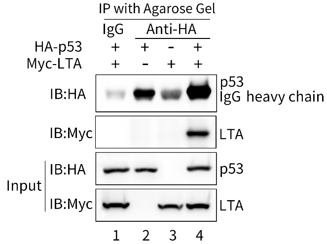
本试剂盒提供三种洗脱方法。根据目的蛋白的结构、生物学功能及后续应用的要求等,本试剂盒提供三种洗脱方法,包括HA多肽、酸性和SDS-PAGE上样缓冲液洗脱液进行洗脱。特别是HA多肽洗脱后不会包含抗体的轻链和重链,可以有效解决免疫沉淀后Western实验中轻链和重链的干扰问题。本试剂盒用于p53和LTA (SV40 Large T antigen)的免疫共沉淀效果参考图2 [4]。

图2.碧云天HA标签蛋白免疫沉淀试剂盒(琼脂糖凝胶法)用于HA-p53和Myc-LTA融合蛋白的免疫共沉淀效果图。293T细胞(人胚肾细胞)单独或共转染pCMV-HA-p53 (D3033)、pCMV-Myc-LTA (D3036)质粒36小时后,使用本试剂盒中的Lysis Buffer裂解。泳道1为Mouse IgG Agarose Gel (小鼠IgG琼脂糖凝胶)免疫沉淀后经本试剂盒提供的SDS-PAGE Sample Loading Buffer洗脱后的样品,该Mouse IgG是正常的小鼠IgG (Normal Mouse IgG),为免疫共沉淀的阴性对照;泳道2、3和4为Anti-HA Agarose Gel免疫沉淀后,经本试剂盒提供的SDS-PAGE Sample Loading Buffer洗脱后的样品,从泳道4可观察到共转染的Myc-LTA与HA-p53可以相互作用。Input即全细胞裂解液(Total cell lysate)。Western印迹成像由BeyoImager™ 600化学发光成像系统完成(EI600)。实际结果会因实验条件、检测仪器等的不同而存在差异,图中数据仅供参考。
本试剂盒提供的Anti-HA Agarose Gel为50%凝胶悬浊液,包装体积为总体积,每毫升中含有0.5ml纯凝胶(沉淀物)。对于常规的免疫沉淀实验,按照每100μl样品使用20μl凝胶悬液,本试剂盒小包装P2206S和中包装P2206M分别可以进行20次和100次样品的免疫沉淀,同时分别可以进行4次和20次阴性对照的免疫沉淀。
包装清单:| 产品编号 | 产品名称 | 包装 |
| P2206S-1 | Lysis Buffer | 50ml |
| P2206S-2 | TBS (10X) | 5ml |
| P2206S-3 | Protease Inhibitor Cocktail (100X) | 0.5ml |
| P2206S-4 | Anti-HA Agarose Gel | 0.4ml |
| P2206S-5 | Mouse IgG Agarose Gel | 80μl |
| P2206S-6 | HA Peptide (25X) | 80μl |
| P2206S-7 | Acid Elution Buffer | 2ml |
| P2206S-8 | Neutralization Buffer | 0.2ml |
| P2206S-9 | SDS-PAGE Sample Loading Buffer (2X) | 6ml |
| - | 说明书 | 1份 |
| 产品编号 | 产品名称 | 包装 |
| P2206M-1 | Lysis Buffer | 250ml |
| P2206M-2 | TBS (10X) | 15ml |
| P2206M-3 | Protease Inhibitor Cocktail (100X) | 2.5ml |
| P2206M-4 | Anti-HA Agarose Gel | 2ml |
| P2206M-5 | Mouse IgG Agarose Gel | 0.4ml |
| P2206M-6 | HA Peptide (25X) | 0.4ml |
| P2206M-7 | Acid Elution Buffer | 10ml |
| P2206M-8 | Neutralization Buffer | 1ml |
| P2206M-9 | SDS-PAGE Sample Loading Buffer (2X) | 3ml |
| - | 说明书 | 1份 |
-20℃保存,一年有效。
注意事项:如果免疫沉淀的目的蛋白涉及磷酸化修饰或者乙酰化修饰,需要自备相应的磷酸酶抑制剂和去乙酰化酶抑制剂。推荐选购碧云天的磷酸酶抑制剂混合物A (50X) (P1081/P1082)和去乙酰化酶抑制剂混合物(100X) (P1112/P1113)。
本试剂盒提供的Lysis Buffer经反复测试,适合很多情况下的免疫沉淀或免疫共沉淀时的样品裂解和后续的洗涤。但由于免疫沉淀或免疫共沉淀蛋白样品的复杂性和特殊性,本Lysis Buffer不一定适合所有免疫沉淀样品的裂解与洗涤。在使用本试剂盒提供的Lysis Buffer效果欠佳的情况下,需要自行对于裂解液和洗涤液进行摸索和调整。此时建议根据文献自行配制裂解液和洗涤液,或尝试碧云天的其它适当裂解液:http://www.beyotime.com/support/lysis-buffer.htm。
Agarose Gel使用前一定要充分重悬,即充分颠倒若干次使混合均匀。
Agarose Gel含有微量的防腐剂,不会影响常规的蛋白或蛋白复合物的纯化和免疫沉淀。但如果后续涉及酶活性测定等防腐剂可能产生干扰的实验,使用前宜先用TBS等适当溶液洗涤凝胶3次,以充分消除防腐剂可能产生的干扰。
在免疫沉淀时,建议设置阳性和阴性对照组(Mouse IgG Agarose Gel)。本试剂盒中提供适量的阴性对照,更多需求,可以订购碧云天的Mouse IgG Agarose (小鼠IgG琼脂糖凝胶) (P2265)。
蛋白样品收集后宜尽快完成纯化工作,并应始终放置在4℃或冰浴,以减缓蛋白降解或变性。
如果离心不能完全除去蛋白样品中的不溶物,可以将样品溶液用0.45μm的滤膜过滤。
本产品仅限于专业人员的科学研究用,不得用于临床诊断或治疗,不得用于食品或药品,不得存放于普通住宅内。
为了您的安全和健康,请穿实验服并戴一次性手套操作。
| Steps | Solution required | Volume per assay |
| Cell lysis and sample preparation | Lysis Buffer with Protease Inhibitor Cocktail | 100μl |
| Preparation of HA Peptide elution buffer and agarose gel | TBS | ~1.6ml |
| Immunoprecipitation | Agarose gel | 20μl |
| Wash (3 times) | Lysis Buffer with Protease Inhibitor Cocktail | 500μl each time |
| Peptide competitive elution (optional) | Peptide solution (1X) | 100μl |
| Acid elution and neutralization (optional) | Acid Elution Buffer | 100μl |
| Neutralization Buffer | 10μl | |
| SDS-PAGE sample loading buffer elution (optional) | SDS-PAGE Sample Loading Buffer (2X) | 20μl |
| Problem | Possible Causes | Solution |
| Very few or no tagged protein exists in the eluate. | Protein is not completely eluted. | Change elution methods. |
| No target protein expressed. | Make sure the protein of interest contains the tagged protein by Western blot or dot blot analyses. | |
| Very low protein expression level. | 1.Use larger volume of cell lysate. 2.Optimize expression conditions to raise the protein expression level. |
|
| Washes are too stringent. | Reduce the time and number of washes. | |
| Incubation times are inadequate. | Increase the incubation time. | |
| Interfering substance is present in sample. | Lysates containing high concentration of DTT, 2-mercaptoethanol, or other reducing agents may destroy antibody function, and must be avoided. | |
| Detection system is inadequate. | If Western blot detection is used: 1.Check primary and secondary antibodies using proper controls to confirm binding and reactivity. 2.Verify that the transfer was adequate by using prestained protein marker or staining the membrane with Ponceau S. 3.Use fresh detection substrate or try a different detection system. |
|
| Background is too high. | Proteins bind nonspecifically to the monoclonal antibody, insufficient washing on agarose gel, or the microcentrifuge tubes. | 1.Pre-clear lysate with Mouse IgG Agarose (P2265) to remove nonspecific binding proteins. 2.After suspending beads for the final wash, transfer entire sample to a clean microcentrifuge tube before centrifugation. |
| Washes are insufficient. | 1.Increase the number of washes. 2.Prolong duration of the washes, incubating each wash for at least 15 minutes. 3.Choose other wash buffers. Increase the salt and/or detergent concentrations in the wash solutions. 4.Centrifuge at lower speed to avoid nonspecific trapping of denatured proteins. |
|
| Multiple protein bands found in the eluate. | The protein is not stable at room temperature. | Purify the target protein at lower temperature, such as 4℃. |
| Protein degradation due to proteases activity during purification process. | Add protease inhibitors to cell lysate. | |
| Non‐specific binding. | 1.Prepare cell lysate again. 2.Add additional wash steps. |


 微信在线咨询


